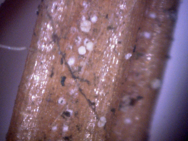
This White spruce in our neighborhood seemed reasonably healthy until this summer when it
started to decline by losing some of its needles.
The last few weeks in fall it started a rapid decline, so I decided to check it out, with the
results shown below.
(As is often the case, details are often hard to see, so click on the pictures
for more detail.)
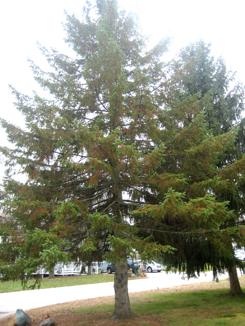
1
This what the entire tree looked like. Unfortunately, the sky was way too
bright, so the decline is hard to see.
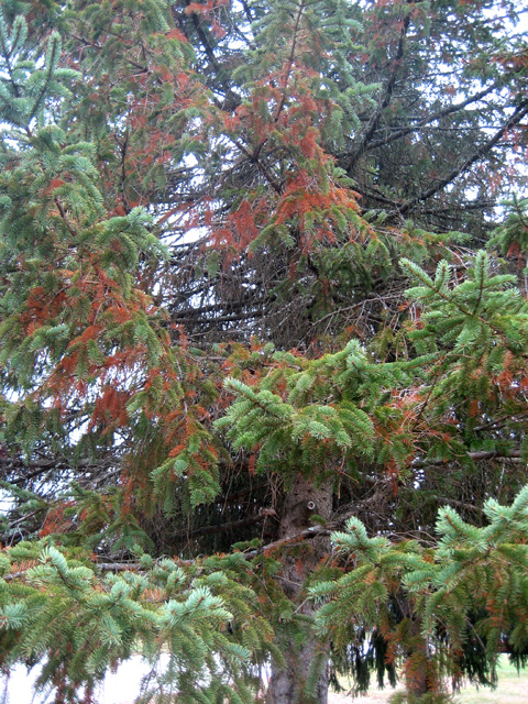
2
This is what the interior of the tree looked like.
You can see that the tree is starting to lose it needles.
Months ago there was none of this dieoff.
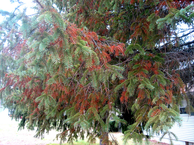
3
This is a view of the lower interior of the tree.
The foliage is already getting fairly sparse, and as the needles are dying, they are taking
on a distinct reddish hue. These reddish needles are very dry and fall off easily when touched.
4
↓
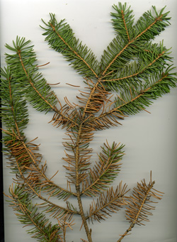
Many of the branches on the tree look just like this one - many dead needles. The
next picture shows a zoom of the area around the branch point indicated by the
red arrow.
5
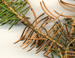
A high resolution picture of the junction between this year's growth, which is green,
and the growth of prior years, which is brown. Note the spores (click the picture
to see them).
6
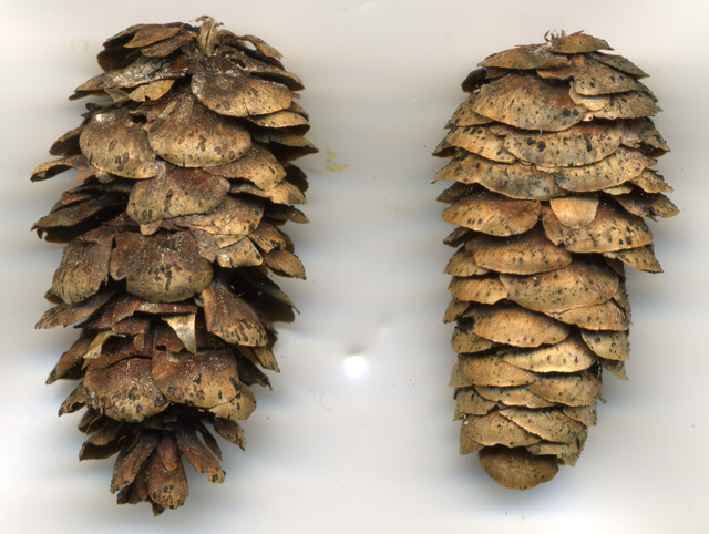
This is a 1200 dpi scan of two pine cones from the tree.
I guessed that the light yellow flecs were white canker spores.
There were far more on the left cone, but both cones have many more at their base.
The above set of 3 pictures was taken using a 1200 dpi scanner. All the following pictures
were taken with a 400x microscope.
7
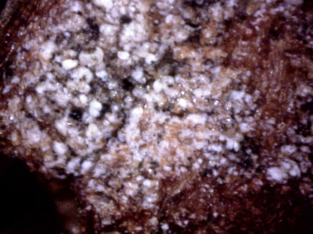
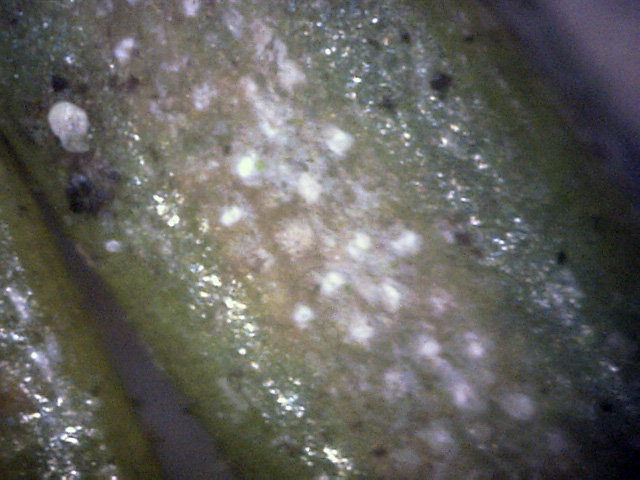
This is a view of a white area on the left pine cone in picture 6.
So indeed these are patches of white canker. White canker seems to love to infect junctions,
or nodes, of a tree.
9
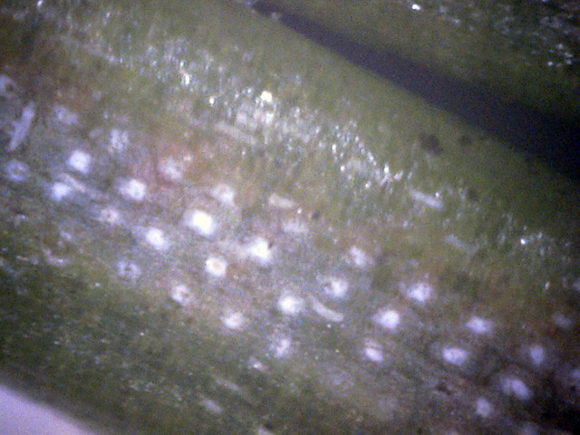
This live needle was only an inch away from dead needles.
The live needles appeared to be only on this year's new growth.
Note the spore on this one, next to a black area on the needle.
It's probably more than a coincidence that this spore is near an area of
white spots - possibly stomata infected with white canker.
10
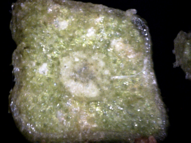
This live needle was also only an inch away from dead needles and was on this year's new growth.
The white spots could be white canker growth.
This is supported by what looks like about 4 hypha that can be seen growing on or just under
the surface of the needle.
11
↓
↓
↓
↓
↑
Cross-section of a live needle. This needle was only inches away from dead needles.
The black arrows point to white canker growths.
White canker is also growing around the pith in the center of the needle.
There is also a hypha of white canker (note the ribbon-like shape) shown by the blue arrow.
 ←
→
←
→
12
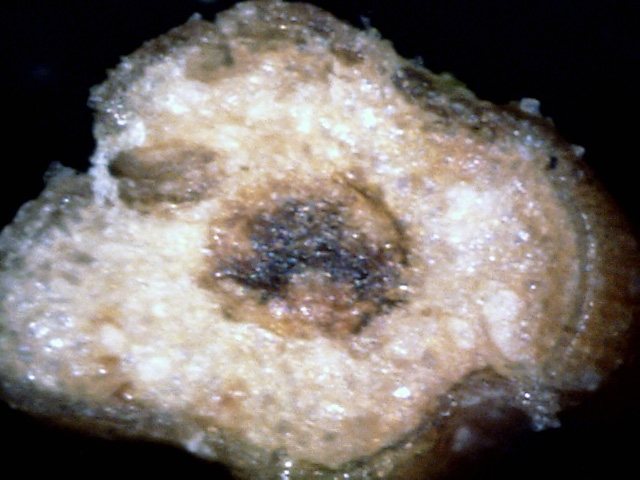
Cross-section of a dead needle. The contrast isn't as clear here, but there is a
large area of white canker on the left, and a large growth of white canker on
the right (black arrows).
13
Cross-section of a dead needle. The center of this needle has pretty much died.
There is so much white canker around the center that it's hard to see the dead
brown surrounding tissue.
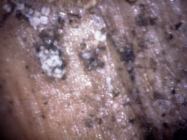
14
These spores are on the surface of the bark. Sometimes they have a tendency to
clump, as they do here.
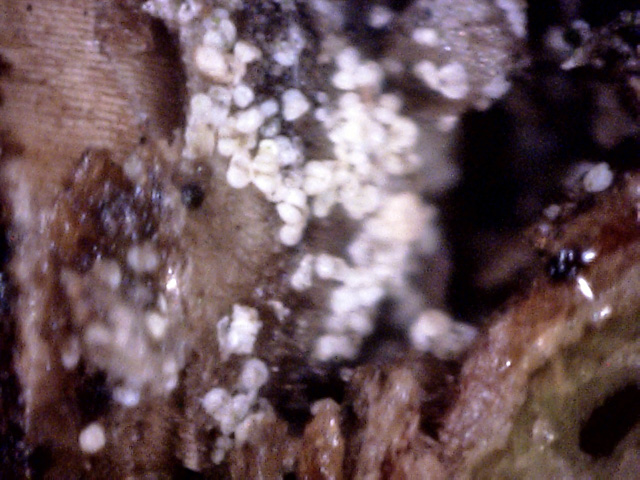
15
This dense spore cluster was located at the junction of two twigs. White canker spores
seem to gravitate to areas near branch junctions.
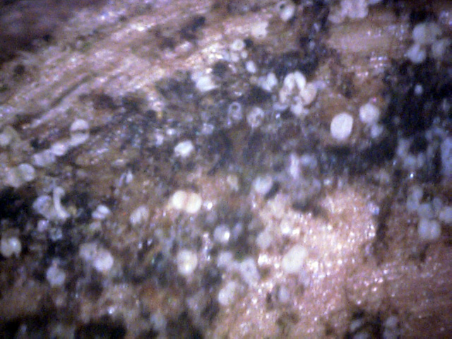
16
There are quite a few spores here, mixed in with a black substance which seems to be spore-related.
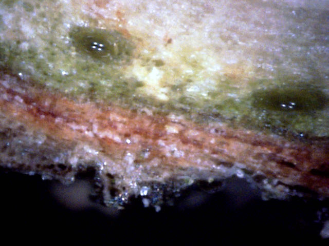
17
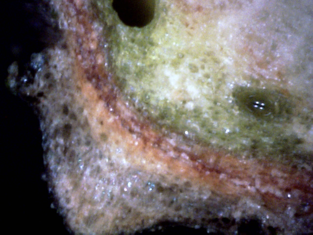
There is canker growth between the two vessels, and quite a bit of canker growth
within the outer bark.
18
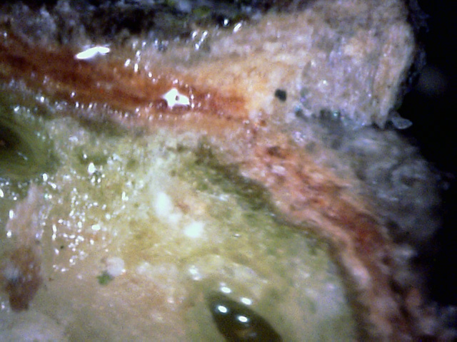
The white canker between the sap vessels has severely infected the nearby outer bark,
pushing it out in the process.
19
The space between the vessels has several spore particles present.
The outer bark surface above it has extensive white canker growth.
In summary, the first severe infection clue was the appearance of orange-brown needles on
the tree. These needles were very dry, fell off easily, and were on all growth over 1 year
old.
(I had seen many spruces like this on a recent trip to Northern Wisconsin. Most
of those trees were dying.) 1200 dpi scans of the branch and pine
cones showed the presence of light yellow particles - possible white canker
spores. Shifting to a 400x microscope, white canker spore patches were confirmed
near the base of the pine cones. While the needle surfaces showed some white
canker, the interiors of the needles showed much more. Areas of the bark surface near branch
junctions had abundant spores. Branch cross-sections showed that, as with other
conifers, white canker is mainly concentrated between the vessels, from where it
pushes out to infect the bark. This white spruce, therefore, definitely
has a moderate white canker infection. But you have to know where to look for
it!
